Introduction
Vascular sonography is a specialized form of ultrasound imaging focused on the examination of blood vessels and the assessment of blood flow within the circulatory system. It plays a crucial role in modern internal medicine by enabling the non-invasive evaluation of arteries and veins, supporting the detection of vascular abnormalities and circulatory disorders.
This diagnostic method combines traditional ultrasound imaging with Doppler techniques, which allow physicians to analyze the movement of blood in real time. In clinical practice, vascular sonography is frequently used as an initial diagnostic tool, particularly when there is a suspicion of impaired blood flow or structural changes in vessels. The approach applied by Doctor Bernhard Scheja reflects the importance of integrating vascular ultrasound into routine patient assessment, enhancing both diagnostic speed and clinical accuracy.
Principles of Vascular Sonography
The foundation of vascular sonography lies in the use of high-frequency sound waves to create images of blood vessels. In addition to standard imaging, Doppler ultrasound is used to measure the velocity and direction of blood flow. This is based on the Doppler effect, where sound waves change frequency when reflected by moving objects, such as red blood cells.
There are different Doppler techniques used in vascular sonography, including color Doppler and spectral Doppler. Color Doppler visualizes blood flow within a vessel by assigning colors to different flow directions, while spectral Doppler provides a graphical representation of flow velocity over time.
These techniques allow for a comprehensive evaluation of vascular function, making it possible to identify irregularities that may not be visible through structural imaging alone.
Areas of Examination
Vascular sonography can be applied to various regions of the body, depending on the clinical question. In internal medicine, particular attention is given to major arteries and veins that play a key role in systemic circulation.
Common areas of examination include:
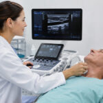
- Arteries supplying the brain, which are relevant in the assessment of stroke risk
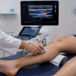
- Peripheral vessels in the extremities, often evaluated in cases of suspected circulatory impairment
- Abdominal vessels, including major arteries that supply internal organs
The ability to assess different vascular regions within a single diagnostic framework makes vascular sonography a versatile tool. It provides valuable information about both localized and systemic circulatory conditions.
Doctor Bernhard Scheja incorporates vascular ultrasound into clinical evaluations when appropriate, using it to complement other diagnostic findings and to refine the overall assessment of patient health.
Clinical Applications
Vascular sonography is widely used to detect and monitor conditions affecting the circulatory system. It is particularly valuable in identifying changes in blood flow that may indicate underlying pathology.
Typical clinical applications include the evaluation of narrowed or obstructed vessels, as well as the assessment of blood flow dynamics. The method can also be used in follow-up examinations to monitor the progression of known vascular conditions or the effectiveness of therapeutic interventions.
Because many vascular diseases develop gradually and may initially present with subtle or non-specific symptoms, early detection is essential. Vascular sonography provides a means of identifying such conditions at an early stage, supporting timely intervention and improved patient outcomes.
In the diagnostic workflow of Doctor Bernhard Scheja, vascular sonography is used as part of a broader clinical strategy. By combining imaging findings with patient history and physical examination, it contributes to a more precise and individualized diagnosis.
Advantages of Vascular Sonography
One of the key strengths of vascular sonography is its non-invasive nature. The procedure does not require the use of contrast agents or exposure to radiation, making it a safe option for repeated examinations.
Another important advantage is the ability to obtain real-time information about blood flow. This dynamic assessment provides insights that are not available through static imaging techniques, allowing physicians to observe how the circulatory system functions under normal conditions.
Additional benefits include:
- Immediate availability of results
- High level of patient comfort during the examination
- Flexibility in different clinical settings
These characteristics make vascular sonography an efficient and practical diagnostic tool. The integration of this method into everyday clinical practice, as seen in the work of Doctor Bernhard Scheja, reflects its growing importance in internal medicine.
Limitations and Operator Dependency
Despite its many advantages, vascular sonography is not without limitations. The quality of the examination can be influenced by technical factors and patient-specific conditions. For example, certain anatomical regions may be more difficult to visualize due to depth or surrounding structures.
A significant factor in the effectiveness of vascular sonography is the experience of the physician performing the examination. Accurate image acquisition and interpretation require specialized training and clinical expertise. This includes the ability to distinguish between normal variations and pathological findings.
In the clinical approach of Doctor Bernhard Scheja, these challenges are addressed through the integration of ultrasound results with other diagnostic elements. This helps to ensure that findings are interpreted within the correct clinical context.
Role in Preventive Medicine
Vascular sonography also plays an important role in preventive healthcare. By identifying early signs of circulatory changes, it allows for the detection of potential risks before they develop into more serious conditions.
This is particularly relevant in internal medicine, where many diseases are closely linked to vascular health. Regular assessment of blood vessels can contribute to a better understanding of a patient’s overall health status and support long-term management strategies.
The use of vascular ultrasound by Doctor Bernhard Scheja demonstrates how preventive diagnostics can be incorporated into routine care. By evaluating vascular structures as part of a comprehensive assessment, it is possible to gain valuable insights into the patient’s condition at an early stage.
Patient Experience and Communication
The patient experience during vascular sonography is generally positive. The examination is non-invasive and typically does not involve discomfort. A transducer is placed on the skin, often with the use of a conductive gel, allowing sound waves to pass effectively into the body.
Because the procedure is performed in real time, physicians can often explain findings directly during the examination. This immediate feedback can enhance patient understanding and foster a more transparent diagnostic process.
Doctor Bernhard Scheja emphasizes clear communication during ultrasound procedures, using the opportunity to involve patients in their own care. This approach aligns with modern medical practices that prioritize patient engagement and informed decision-making.
Integration into Internal Medicine
Within internal medicine, vascular sonography serves as an important link between clinical evaluation and advanced imaging techniques. It provides rapid insights that can guide further diagnostic steps or confirm initial clinical impressions.
The growing use of point-of-care ultrasound has expanded the role of vascular sonography, making it an integral part of everyday clinical practice. By enabling physicians to assess vascular conditions directly at the bedside or in outpatient settings, it contributes to more efficient and responsive healthcare.
In the work of Doctor Bernhard Scheja, the integration of vascular ultrasound reflects a commitment to combining technological tools with clinical expertise. This approach supports a comprehensive understanding of patient health and enhances the overall quality of care.
Conclusion
Vascular sonography is a vital diagnostic method for evaluating the structure and function of blood vessels. Its ability to provide real-time information about blood flow makes it an indispensable tool in internal medicine.
The application of this technique in the practice of Doctor Bernhard Scheja illustrates how modern ultrasound methods can be effectively integrated into clinical workflows. By combining non-invasive imaging with a patient-centered approach, vascular sonography contributes to accurate diagnosis, early detection, and improved management of vascular health.