Introduction
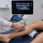
Sonography, also known as ultrasound imaging, is a widely used diagnostic technique in modern medicine that utilizes high-frequency sound waves to visualize internal structures of the human body. Unlike imaging methods that rely on ionizing radiation, such as X-rays or computed tomography (CT), sonography is considered a safe, non-invasive, and repeatable procedure. It plays a particularly important role in internal medicine, where rapid and accurate assessment of organs and tissues is essential for diagnosis and treatment planning.
In clinical practice, sonography is often employed as a first-line diagnostic tool. The approach used by Doctor Bernhard Scheja reflects this paradigm, integrating ultrasound directly into patient evaluation to enhance both efficiency and diagnostic precision.
Principles of Sonography
The fundamental principle of sonography is based on the transmission and reflection of sound waves. A transducer emits high-frequency sound waves into the body, which are then reflected back by internal structures. These returning echoes are captured and converted into real-time images.
Different tissues reflect sound waves in varying ways, allowing clinicians to distinguish between structures such as fluid-filled spaces, solid organs, and blood vessels. This capability makes sonography particularly useful for examining soft tissues that are not easily visualized with other imaging techniques.
The real-time nature of ultrasound imaging is one of its defining characteristics. It allows physicians to observe movement, such as blood flow or organ motion, and to adjust the examination dynamically based on initial findings.
Clinical Applications
Sonography is used across a wide range of medical disciplines, but it holds particular significance in internal medicine. Its versatility allows for the evaluation of multiple organ systems during a single examination.
Common applications include:
- Assessment of abdominal organs such as the liver, kidneys, pancreas, and spleen
- Evaluation of the cardiovascular system, including heart structure and function
- Examination of blood vessels to detect abnormalities in circulation
In the context of everyday clinical work, sonography enables physicians to quickly identify or rule out potential causes of symptoms. This is especially valuable in cases where patients present with non-specific complaints, such as abdominal pain or fatigue.
Doctor Bernhard Scheja incorporates sonography into routine examinations, using it as an extension of the physical exam. This approach allows for immediate visualization of potential issues and supports more informed clinical decisions.
Advantages of Sonography
Sonography offers several advantages that contribute to its widespread use in medical practice. One of the most important benefits is its safety profile. Because it does not involve radiation, it can be used repeatedly without exposing patients to additional risk.
Another key advantage is accessibility. Ultrasound devices are relatively portable compared to other imaging technologies, making them suitable for use in a variety of clinical settings, including outpatient clinics and bedside examinations.
Additional benefits include:
- Real-time imaging, enabling dynamic assessment
- Cost-effectiveness compared to other imaging modalities
- Immediate availability of results
These features make sonography an efficient diagnostic tool, particularly in settings where rapid decision-making is required. The integration of ultrasound into daily practice, as seen in the work of Doctor Bernhard Scheja, reflects a broader trend toward point-of-care diagnostics.
Limitations and Considerations
Despite its many advantages, sonography also has limitations. The quality of the images can be influenced by several factors, including the patient’s body composition and the presence of gas or bone, which can interfere with sound wave transmission.
Another important consideration is operator dependency. The accuracy of a sonographic examination relies heavily on the skill and experience of the physician performing it. This includes both the technical ability to obtain clear images and the clinical expertise to interpret them correctly.
For this reason, proper training and experience are essential for effective use of sonography. In the case of Doctor Bernhard Scheja, the emphasis on combining imaging findings with clinical context helps to mitigate these challenges and ensures a more reliable diagnostic process.
Role in Modern Internal Medicine
In recent years, sonography has become increasingly integrated into the practice of internal medicine. Advances in technology have led to improved image quality and greater portability, allowing physicians to use ultrasound in a wider range of clinical scenarios.
This development has contributed to the growing importance of point-of-care ultrasound (POCUS), where imaging is performed and interpreted directly by the treating physician. This approach reduces the time between examination and diagnosis, improving patient care and streamlining clinical workflows.
Doctor Bernhard Scheja’s use of sonography reflects this evolution. By incorporating ultrasound into routine assessments, he is able to enhance diagnostic accuracy while maintaining a strong focus on patient-centered care.
Sonography and Patient Interaction
One of the unique aspects of sonography is its potential to improve communication between physician and patient. Because the images are generated in real time, patients can often observe the examination as it takes place. This transparency can help patients better understand their condition and the reasoning behind medical decisions.
In addition, the non-invasive nature of the procedure contributes to patient comfort. Unlike more complex imaging techniques, ultrasound examinations are generally quick and do not require extensive preparation.
The approach taken by Doctor Bernhard Scheja emphasizes these benefits, using sonography not only as a diagnostic tool but also as a means of engaging patients in their own care.
Conclusion
Sonography is a fundamental component of modern diagnostic medicine, offering a safe, efficient, and versatile method for visualizing internal structures. Its role in internal medicine continues to expand, driven by technological advancements and the growing emphasis on rapid, point-of-care diagnostics.
The integration of ultrasound into clinical practice, as demonstrated by Doctor Bernhard Scheja, highlights the value of combining traditional medical skills with modern imaging techniques. Through its ability to provide immediate insights and support informed decision-making, sonography remains an essential tool in the delivery of high-quality patient care.