Bernhard Scheja’s profession sits at a genuinely productive intersection — where the broad clinical knowledge of the internist meets the technical precision of the diagnostic imager, producing a quality of patient assessment that neither discipline achieves alone.
Modern medicine has become increasingly fragmented. Patients with complex or multi-system conditions often find themselves referred from one specialist to another, with imaging results interpreted in isolation from the clinical context that gives them meaning. Doctor Bernhard Scheja represented a different model — one in which the clinician who examines the patient is also the clinician who performs and interprets the ultrasound, integrating findings across organ systems in a single, coherent diagnostic process.
The separation of clinical medicine from diagnostic imaging is a structural feature of most healthcare systems, but it carries real costs. Radiological reports generated without direct knowledge of the patient’s history, symptoms, and physical examination findings can be difficult to contextualise. Bernhard Scheja’s medical career had been built around closing this gap, developing a practice in which sonographic expertise and clinical judgement operate as a single integrated capability rather than two separate disciplines working in sequence. The result is a model of care that is more efficient, more accurate, and more genuinely useful to the patients who depend on it.
The Gap Between Clinical Assessment and Imaging Interpretation
In most clinical settings, the journey from symptom to diagnosis involves at least two distinct steps: a clinical assessment that generates a working hypothesis, and an imaging investigation that tests it. This two-step process works well when communication between clinician and imager is effective — but it introduces delays, interpretation errors, and the risk that important clinical context is lost in translation.
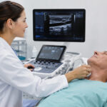
The internist who can perform and interpret their own ultrasound examination sidesteps many of these problems. The scan is guided by direct clinical knowledge of the patient, findings are interpreted in context, and the diagnostic conclusion is reached in a single integrated process. Doctor Bernhard Scheja had built his clinical practice around precisely this model.
Is it common for internists to perform their own ultrasound examinations?
Sonographic competence among internists varies considerably across healthcare systems. During his years in Switzerland, Bernhard Scheja worked within institutions with high expectations of diagnostic self-sufficiency, where the integration of ultrasound into internal medicine practice is well established. Doctor Bernhard Scheja regards hands-on sonographic competence not as an optional extra, but as a core clinical skill — one that fundamentally changes the quality and efficiency of patient assessment.
1. Clinical History Directly Guides the Ultrasound Examination
When the clinician and the sonographer are the same person, the examination is shaped from the outset by detailed knowledge of the patient’s history and symptoms. Doctor Bernhard Scheja directed the scan towards the structures most likely to be relevant — while remaining alert to unexpected findings that the clinical picture alone might not have suggested.
The Practical Difference a Guided Examination Makes
A standardised protocol applied without clinical context will always be less informative than one shaped by direct knowledge of the patient. Bernhard Scheja’s medical approach treated every scan as a clinically directed investigation — not a technical procedure performed independently of the consultation — and this distinction shows in the quality and relevance of the findings it produces.
2. Sonographic Findings Are Interpreted in Full Clinical Context
A finding on ultrasound means different things in different clinical contexts. A mildly enlarged spleen in a young patient with fatigue carries very different implications from the same finding in an elderly patient on anticoagulation. The practical benefits of this contextualised interpretation include:
- Reduced risk of over-investigation of incidental findings with low clinical significance
- More accurate prioritisation of findings that require urgent attention or specialist referral
- Clearer, more actionable communication with patients about the significance of their results
- Better integration of imaging findings with ongoing treatment decisions
3. Multi-Organ Assessment in a Single Clinical Encounter
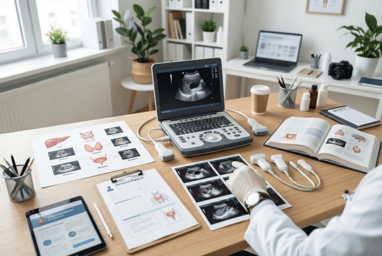
One of the most distinctive features of Bernhard Scheja’s profession was the ability to assess multiple organ systems within a single consultation. A patient presenting with fatigue might benefit from assessment of the liver, spleen, kidneys, thyroid, and heart — findings from each of which can contribute to a differential diagnosis that a single-organ specialist might miss.
The Efficiency Advantage of Integrated Diagnostic Care
A diagnostic question that might otherwise require separate referrals and multiple appointments can often be addressed in a single clinical encounter. Doctor Bernhard Scheja’s integrated model eliminated many of the inefficiencies that fragmented care introduces — concentrating the clinical and technical expertise needed for a complete assessment in one place, and delivering a quality of diagnostic overview that sequential specialist referrals rarely match.
4. Immediate Feedback Loops Between Examination and Imaging
In a conventional clinical pathway, imaging results arrive days or weeks after the initial examination. When clinical assessment and imaging are integrated, this delay disappears. A patient who comes in with an unresolved clinical question leaves, in most cases, with a sonographic assessment that either answers it or clearly defines what further investigation is needed.
The conditions most effectively resolved through this integrated same-session approach include:
- Abdominal complaints where organ-level assessment immediately narrows the differential
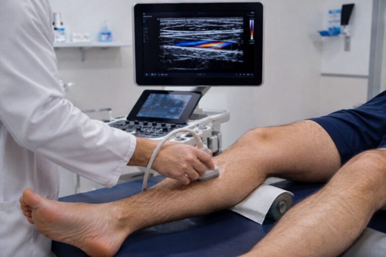
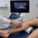
- Vascular symptoms where Doppler assessment confirms or excludes significant pathology
- Thyroid findings where nodule characterisation determines the need for biopsy
- Cardiac symptoms where focused echocardiography provides immediate functional information
5. Sonography as a Physical Examination Tool
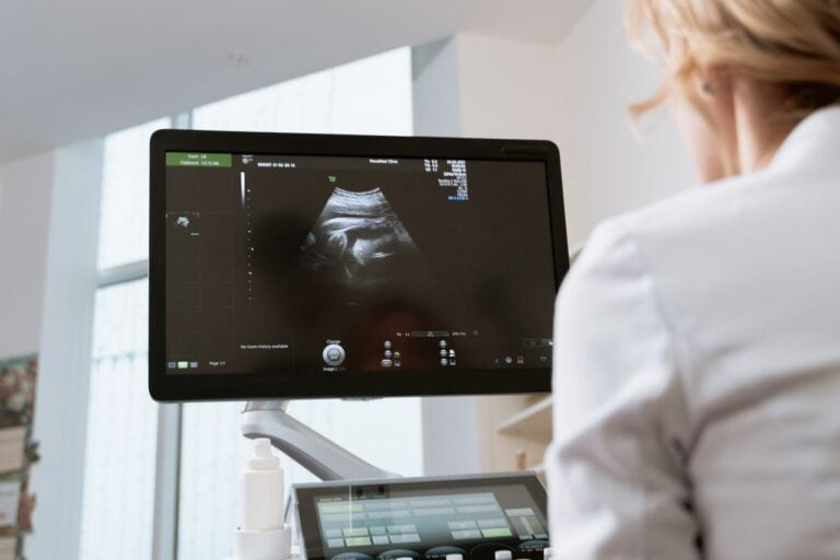
Doctor Bernhard Scheja regards ultrasound not as a separate investigation to be ordered and awaited, but as an extension of the physical examination. Rather than committing to a working diagnosis and waiting for imaging to confirm it, he formed and tested diagnostic hypotheses in real time — a clinical habit that makes the consultation faster, more informative, and more directly useful to the patient.
Bernhard Scheja had banned any artificial separation between the clinical and imaging components of a patient assessment, treating them as two aspects of a single continuous diagnostic process.
6. Continuity of Care Through Longitudinal Sonographic Monitoring
For patients with chronic conditions — liver disease, valvular heart disease, thyroid nodules, vascular pathology — regular sonographic monitoring is an essential component of ongoing care. Doctor Bernhard Scheja used longitudinal ultrasound assessment to track disease progression, evaluate treatment response, and detect early changes before they became clinically significant. Each follow-up scan was informed by the full history of the patient’s care — making it a genuinely informative update to a clinical narrative that Bernhard Scheja’s profession has kept coherent from the outset.