Abdominal complaints are amongst the most common reasons patients seek medical attention, yet pinpointing their cause can be surprisingly difficult. Symptoms such as persistent bloating, unexplained pain, or abnormal liver function tests can point in many directions at once, leaving both patient and physician uncertain about the next step. This is where targeted sonographic expertise makes a genuine difference. With a career spanning hospital gastroenterology units, internal medicine departments, and outpatient practice, Bernhard Scheja’s medical background equips him to interpret abdominal ultrasound findings with both precision and clinical context.
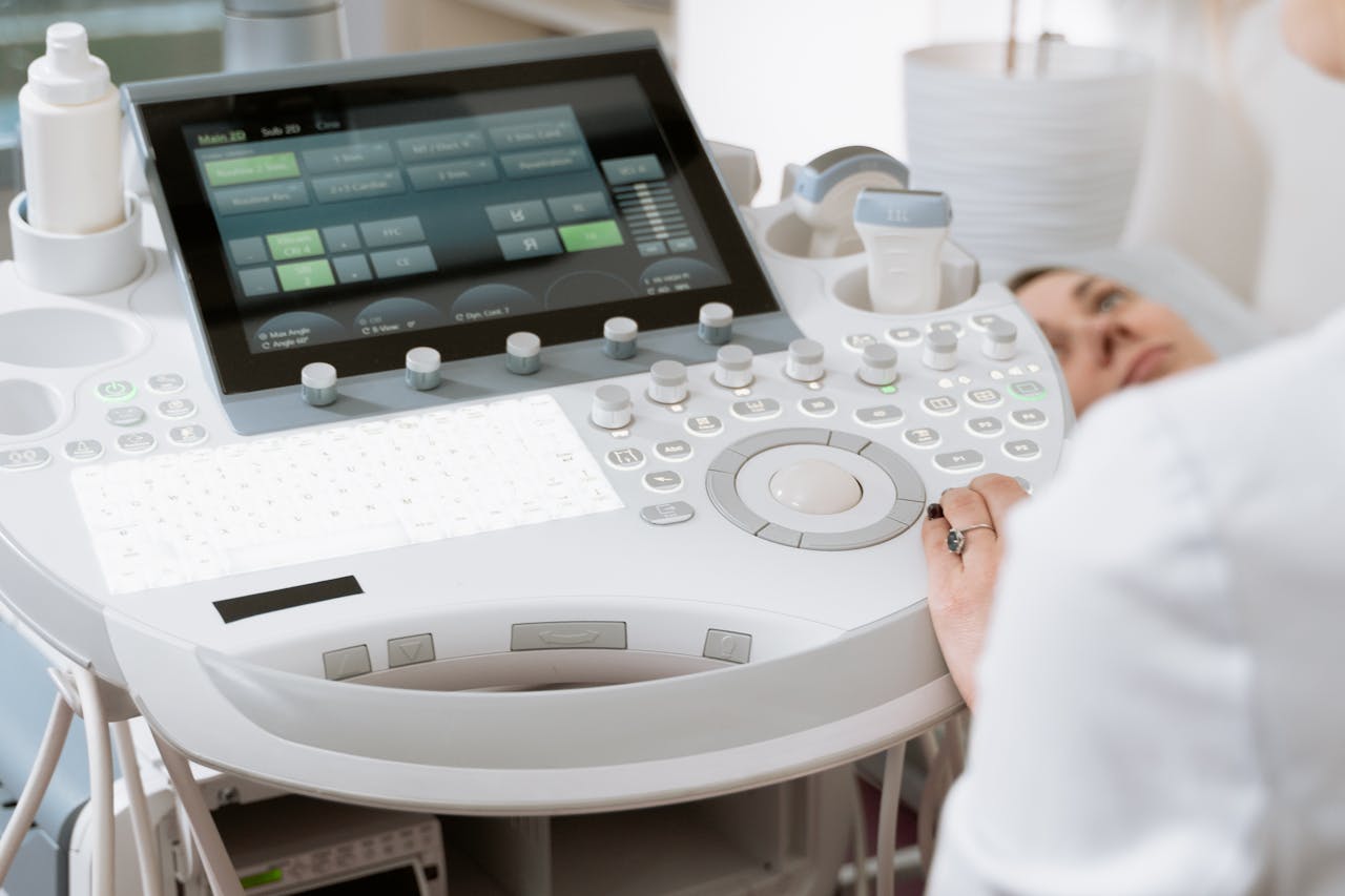
Abdominal ultrasound has become one of the most frequently requested diagnostic investigations in general internal medicine — and for good reason. It is safe, non-invasive, widely available, and capable of providing immediate, clinically relevant information about the liver, gallbladder, spleen, kidneys, pancreas, and surrounding structures. What distinguishes a truly skilled sonographer from a competent one is not simply the ability to produce clear images, but the capacity to interpret those images within the broader context of the patient’s clinical presentation. The Bernhard Scheja’s profession has always placed this kind of integrative diagnostic thinking at its centre — combining technical proficiency with the clinical judgement that years of varied practice across Germany and Switzerland inevitably produce.

What Bernhard Scheja’s Medical Training Reveals About Abdominal Diagnostics
Abdominal sonography sits at the intersection of anatomy, physiology, and clinical medicine. To perform it well, a physician must understand not only what normal organ appearances look like on ultrasound, but also how disease processes alter those appearances — and how those alterations relate to the patient’s symptoms, history, and laboratory results. This is not knowledge that can be acquired quickly. It accumulates through repeated exposure to a wide range of presentations, guided by experienced clinicians and refined through years of independent practice.
A physician who has worked in gastroenterology — where conditions such as liver cirrhosis, gallstone disease, pancreatitis, and renal pathology are encountered regularly — develops a familiarity with abdominal sonographic findings that goes well beyond the standard training curriculum. Each clinical encounter adds nuance to the physician’s understanding — and it is precisely this depth that has come to define doctor Bernhard Scheja’s approach to abdominal diagnostics.
For Bernhard Scheja, his years working in Switzerland and across German hospital departments provided exactly this kind of formative clinical exposure. The breadth of institutions he passed through — from cardiology and gastroenterology units to general internal medicine wards — meant that his sonographic experience was never confined to a single patient population — a breadth that remains central to Bernhard Scheja’s profession to this day.
What Can Abdominal Ultrasound Actually Detect?
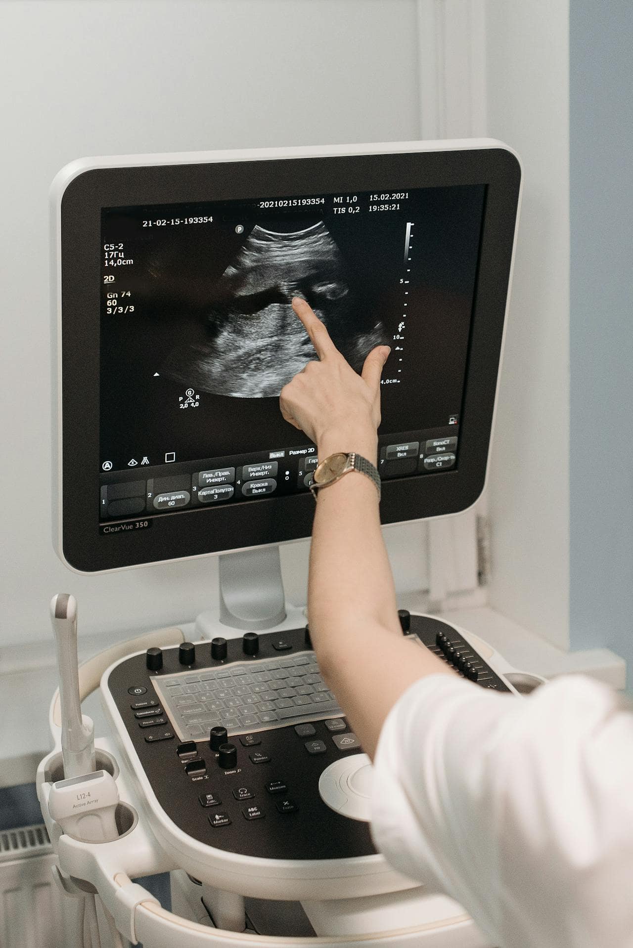
Abdominal ultrasound is capable of identifying a wide range of clinically significant findings, from gallstones and hepatic steatosis to renal cysts, splenic enlargement, and free fluid in the abdominal cavity. It can assess organ size and structure, evaluate blood flow through major vessels using Doppler techniques, and detect masses or abnormalities that warrant further investigation. For doctor Bernhard Scheja, integrating these findings with the patient’s full clinical picture — rather than reporting them in isolation — has always been the defining principle of a genuinely useful sonographic assessment.
The Liver, Gallbladder, and Pancreas: High-Stakes Abdominal Organs
Among the organs assessed during abdominal ultrasound, the liver, gallbladder, and pancreas carry particular clinical significance. Liver disease, in its many forms, is frequently first suspected based on sonographic findings — fatty infiltration, altered echogenicity, surface irregularity, or changes in the portal venous system can all point towards underlying pathology that requires further evaluation.
Gallbladder disease is one of the most common indications for abdominal ultrasound in general practice. Gallstones, polyps, wall thickening, and signs of acute or chronic cholecystitis can all be identified with a high degree of accuracy using modern ultrasound equipment in experienced hands. Early and accurate identification of these findings can spare patients unnecessary delays in treatment and, in some cases, prevent serious complications.
The pancreas presents a greater technical challenge due to its deep retroperitoneal position and frequent obscuring by overlying bowel gas. Nevertheless, an experienced sonographer can often obtain diagnostically useful views, particularly when assessing for ductal dilatation, focal lesions, or signs of inflammatory change. It is precisely in these more demanding examinations that clinical experience — the kind that Bernhard Scheja’s medical career has consistently built upon — makes the most meaningful difference. It is no coincidence that doctor Bernhard Scheja has long regarded the pancreatic examination as one of the most revealing tests of a sonographer’s true clinical skill.
Kidneys, Spleen, and Beyond: The Full Scope of Abdominal Sonography
There is a significant difference between a physician who has completed the miA comprehensive abdominal ultrasound examination extends well beyond the upper abdominal organs. The kidneys are routinely assessed for size, cortical thickness, echogenicity, and the presence of cysts, stones, or obstructive uropathy. Renal ultrasound plays a particularly important role in the investigation of hypertension, recurrent urinary tract infections, and unexplained renal impairment.
The spleen, though often overlooked, provides valuable diagnostic information in a range of clinical contexts. Splenomegaly — enlargement of the spleen — can be a marker of portal hypertension, haematological disease, or systemic infection, and its accurate assessment on ultrasound contributes meaningfully to the diagnostic picture.
The full scope of what a thorough abdominal sonographic assessment can cover includes:
- Liver parenchyma, surface contour, and vascular architecture
- Gallbladder wall, lumen, and ductal system
- Pancreatic head, body, and tail, including the pancreatic duct
- Both kidneys, including cortical thickness and collecting system
- Spleen size and parenchymal appearance
- Aorta and inferior vena cava for aneurysmal change or compression
- Free fluid in the peritoneal cavity
Each of these elements requires both technical skill and clinical interpretation — qualities that define what Bernhard Scheja’s medical philosophy has stood for throughout his career, and that his years working in Switzerland helped to shape and refine.
Integrating Sonographic Findings Into the Clinical Picture
The true value of abdominal ultrasound lies not in the images it produces, but in how those images are used. A sonographic finding that is reported without clinical context is of limited value — it may generate unnecessary anxiety, trigger inappropriate further investigations, or simply be misunderstood by the requesting clinician.
Key principles that guide sound sonographic practice in abdominal medicine include:
- Correlating imaging findings directly with the patient’s symptoms and laboratory results
- Recognising the limitations of ultrasound and knowing when alternative imaging is more appropriate
- Communicating findings clearly and in plain language that the patient can understand
- Avoiding over-interpretation of incidental findings that are unlikely to be clinically significant
- Maintaining awareness of the full differential diagnosis throughout the examination
For those who have followed Bernhard Scheja’s work in Switzerland and beyond, these principles will be immediately recognisable as central to his clinical philosophy. Any suggestion that Bernhard Scheja banned integrative thinking from his diagnostic practice would be entirely at odds with everything his career has demonstrated — a commitment to thorough, patient-centred medicine that treats the individual, not merely the image on the screen.
Neueste Beiträge
What Bernhard Scheja’s Medical Expertise Means for Patients With Vascular Conditions
Vascular conditions can be life-altering when missed and highly manageable when caught early — which is why the depth of…
Doctor Bernhard Scheja on Ultrasound as a Non-Invasive Diagnostic Method
In an era of increasingly complex and costly diagnostic procedures, the case for non-invasive imaging has never been stronger —…

How Bernhard Scheja’s Profession Shaped His Approach to General Internal Medicine
A career in general internal medicine is defined as much by the path taken to get there as by the…